Table of Contents
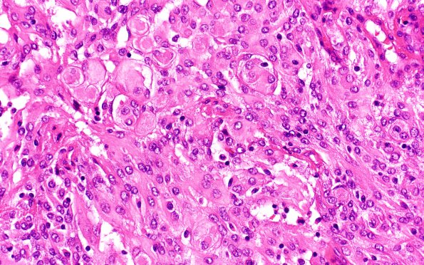
Washington University Experience | NEOPLASMS (MENINGIOMA) | Anaplastic | 13C6 Meningioma, anaplastic (Case 13) H&E 40X
H&E stained sections of the resection material show a high grade meningioma with large, predominantly central, areas of geographic necrosis that comprise approximately 80% of the specimen. Mitotic figures are frequent, appearing at variable density that focally reaches 21/10HPF. Brain invasion is also identified focally. Sections of the dura show areas of tumor invasion. Comment: This histomorphological pattern is diagnostic for anaplastic meningioma with focal rhabdoid/chordoid features, WHO grade III. The grade III diagnosis is established by mitotic figures in excess of 20/HPF. Rhabdoid cytoarchitecture substantiates a grade III diagnosis only if it comprises the majority of the tumor, which is not true in this case; "with rhabdoid features" is appended to the diagnosis because this histological pattern is uncommon and may be relevant to diagnosis in the event of recurrence. FISH analysis was performed to evaluate for chromosome 9p21 loss (CDKN2A/p16 locus); in this case, CDKN2A loss was detected within this tumor. Deletion of 9p21, found in most anaplastic meningiomas, is associated with relatively unfavorable prognosis.

