Table of Contents
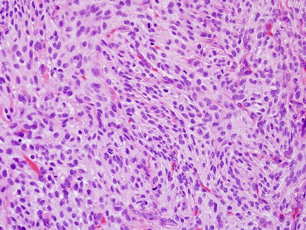
Washington University Experience | NEOPLASMS (MENINGIOMA) | Anaplastic | 14B1 Meningioma, anaplastic (Case 14) H&E 5.jpg
14B1-3 The tumor specimen shows multiple fragments of a dura based, meningothelial cell neoplasm with 'sheet-like' patternless growth, hypercellularity, small cell change, and multifocal necrosis. In a randomly chosen area, mitotic figures were found at 35 mitoses/HPF. The tumor cells have a spindled appearance and are largely without distinct cell borders. The tumor nuclei show slight pleomorphism, are oval to spindled, have finely speckled chromatin, and some have prominent nucleoli.

