Table of Contents
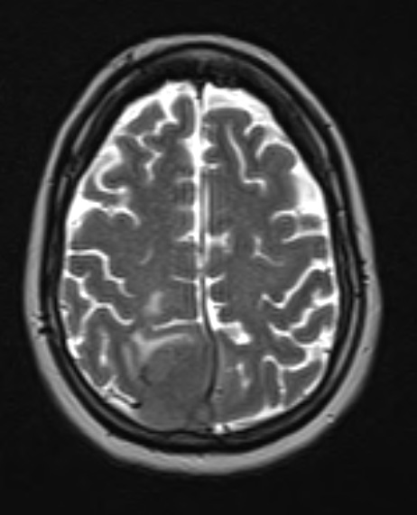
Washington University Experience | NEOPLASMS (MENINGIOMA) | Anaplastic | 15A3 Meningioma, anaplastic (Case 15) T2 W 1 - Copy
Brain MRI presented as T2-weighted scan showed a 4.8 cm post-contrast enhancing parafalcine mass with invasion into the superior sagittal sinus and right parietal bone.

