Table of Contents
Washington University Experience | NEOPLASMS (MENINGIOMA) | Anaplastic | 18A1 Meningioma, anaplastic (Case 18) 2.jpg
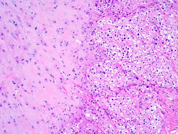
Case 18 History ---- The patient is a 77 year old woman with a 5.1 x 2.0 cm irregular extra-axial mass along the left temporal convexity. The radiologic differential diagnosis included atypical meningioma and a dural metastasis. 18A1-3 The tumor has a heterogeneous histologic appearance. In some areas, the tumor is composed predominantly of sheets of clear cells with round to oval nuclei, prominent nucleoli, and abundant clear to eosinophilic cytoplasm. Only vague whorls are seen in these areas. Other areas have a sarcoma-like appearance with islands of hyalinized and cartilaginous tissue containing large, bizarre nuclei, some of which have frankly malignant cytologic features. Some of the tumor cells are located within lacunar spaces set within a myxochondroid background, resembling chondrosarcoma. Mitotic figures are relatively uncommon, although there are foci of tumor necrosis evident. No brain parenchyma is seen.

