Table of Contents
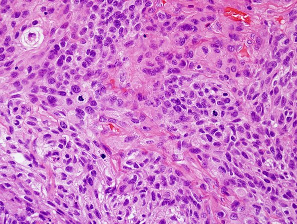
Washington University Experience | NEOPLASMS (MENINGIOMA) | Anaplastic | 19B3 Meningioma, anaplastic (Case 19) H&E 4.jpg
19B3-5 In many areas, the tumor tissue is histologically consistent with meningothelial meningioma; epithelioid and spindled tumor cells with oval nuclei, eosinophilic cytoplasm, indistinct cell borders are arranged in whorls and fascicles. However, these areas also show numerous mitotic figures, ranging up to 40/10HPF, as well as extensive spontaneous necrosis and hypercellularity. In other areas, the tumor exhibits sheeted architecture, prominent nucleoli, regional chordoid, clear cell, and carcinomatous features, and widespread brain invasion.

