Table of Contents
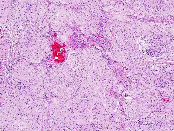
Washington University Experience | NEOPLASMS (MENINGIOMA) | Anaplastic | 21A1 Meningioma, anaplastic (Case 21) H&E 3.jpg
Case 21 History ---- The patient was a 19 year old man who complained of headache on the left side of the face, swelling and a pressure-type sensation for approximately 2 months. CT imaging showed a mixed density mass and surrounding edema at the left middle cranial fossa, with mass effect on the left lateral ventricle. The patient underwent pre-operative arterial embolization, followed by resection. ---- 21A1-3 This is a neoplastic proliferation of meningothelial cells arranged in whorls and ill-defined fascicles. The cells are characterized by moderate amounts of eosinophilic cytoplasm, and predominantly ovoid-shaped nuclei with coarse, vesicular chromatin, occasional multinucleation, and occasional nuclear clearing and intracytoplasmic nuclear inclusions. The tumor also shows focal hypercellularity and areas suggestive of small cell change. Occasional macronucleoli are observed. Focal brain invasion is identified. Multiple foci of necrosis are seen; however, the patient had undergone embolization, and the significance of this necrosis is unclear. Finally, the tumor displays a markedly elevated mitotic index, ranging up to 21-23 mitotic figures/10HPF.

