Table of Contents
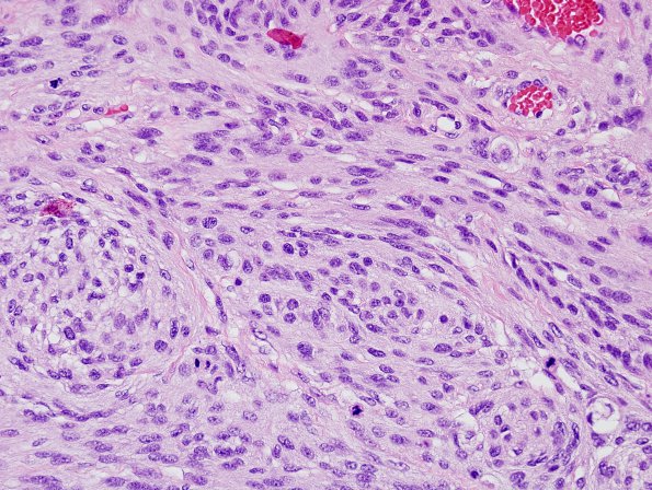
Washington University Experience | NEOPLASMS (MENINGIOMA) | Anaplastic | 21A3 Meningioma, anaplastic (Case 21) H&E 4.jpg
This is a neoplastic proliferation of meningothelial cells arranged in whorls and ill-defined fascicles. The tumor also shows focal hypercellularity and areas suggestive of small cell change. Multiple foci of necrosis are seen; however, the patient had undergone embolization, and the significance of this necrosis is unclear. Finally, the tumor displays a markedly elevated mitotic index, ranging up to 21-23 mitotic figures/10HPF.

