Table of Contents
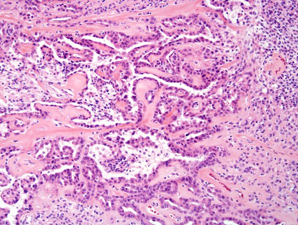
Washington University Experience | NEOPLASMS (MENINGIOMA) | Anaplastic | 24A Meningioma, WHO III w metaplasia (Case 24)
Case 24 History ---- The patient was a 50 y/o female originally diagnosed with an atypical meningioma (WHO grade II) of the cerebellopontine angle in 2000. Microscopic sections from the initial 2000 resection revealed an atypical meningioma, WHO grade II. Whorls of tumor cells are identified but psammoma bodies are absent. Necrosis and brain invasion are also absent, but scattered mitoses are seen and number 4/10 high power fields. Tumor nuclei are oval and bear bland chromatin and indistinct nucleoli; associated cytoplasm is eosinophilic and ill-defined. There has since been a clinical recurrence at the site of prior resection. ---- 24A Microscopic sections from the 2007 resection reveal an anaplastic meningioma now displaying both glandular metaplasia and focal secretory features. Tumor cell density is greater than that of the 2000 primary. Scattered throughout the tumor are true glands and numerous signet ring-like cells. Well differentiated meningiomatous features are not conspicuous and the glandular structures resemble adenocarcinoma. Occasionally, micropapillary structures emanate from the glandular epithelium. Pseudopsammoma bodies are focally identified. Non-glandular areas are populated by sheets and clusters of tumor cells that create a vague lobularity at low power magnification. There are focal 'small cells'. Mitoses are numerous and number 18/10HPF. Tumor cells are more atypical than in the 2000 specimen. Tumor nuclei are oval/angulate in shape and contain open chromatin with prominent nucleoli. Tumor cell cytoplasm is again eosinophilic and ill-defined. Notably, the tumor cells comprising the glands are cytologically similar to those in the non-glandular areas. Necrosis and brain invasion are absent.

