Table of Contents
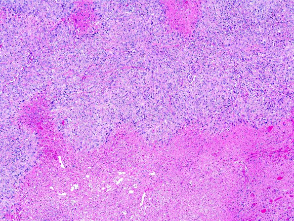
Washington University Experience | NEOPLASMS (MENINGIOMA) | Anaplastic | 25A1 Meningioma, anaplastic (Case 25) 1.jpg
Case 25 History ---- The patient was a 72 year old man who presented with vertigo. Imaging studies revealed a left cerebral cyst with diffuse enhancement of the wall and a region of nodular enhancement superiorly. The patient also had had a long standing arachnoid cyst within this same region. ---- 25A1-3 The specimen consists of a thick plaque-like, extensively necrotic neoplasm with moderate cellularity. The areas of viable tumor are arranged in sheets and fascicles of epithelioid and spindled cells with abundant eosinophilic cytoplasm. Tumor nuclei are mostly oval with vesicular chromatin and prominent nucleoli. The mitotic index is increased and occasional atypical mitotic figures are seen. Focally, there are areas of perivascular hyalinization, vaguely resembling whorls. Other parts of the tumor similarly show variable production of collagen.

