Table of Contents
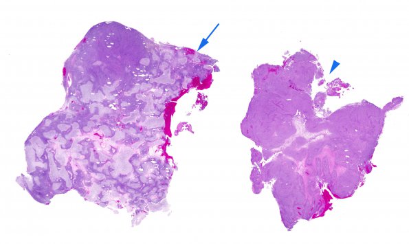
Washington University Experience | NEOPLASMS (MENINGIOMA) | Anaplastic | 26A1 Meningioma, anaplastic & Chondrosarc (Case 26) H&E WM copy
Case 26 History ---- The patient was a 71-year-old woman who experienced seizures in 2012, and was found to have a tumor in the left middle cranial fossa. The patient underwent resection in 02/2012 at the referring institution. In 09/2013, she underwent re-resection for recurrence. The patient was then treated with fractionated radiation therapy as well as concurrent Temodar. In 05/2014 and 07/2014, she suffered seizures, the latter associated with new aphasia and right-sided hemiparesis. Magnetic resonance imaging performed at an outside institution in 05/2014 showed a 1 x 0.6 cm nodular/lobular area of enhancement associated with the resection bed. Repeat imaging in 08/2014 performed at BJH showed an interval increase in the nodular/lobular area of enhancement. Radiological impression: Radiation necrosis and/or tumor recurrence. The pathology materials shown are from the 2013 resection. ---- 26A1-3 The majority of the tumor appears sarcomatous (26A1, arrowhead), with dense arrays of small, spindled cells arranged into broad fascicles and focal storiform arrangements, reminiscent of fibrosarcoma. Foci of geographic necrosis are widespread, in some areas sculpting viable tumor into large perivascular lobules. There is significant variation in the histological appearance of this tumor from place to place. Mitoses exceed 20/10HPF.

