Table of Contents
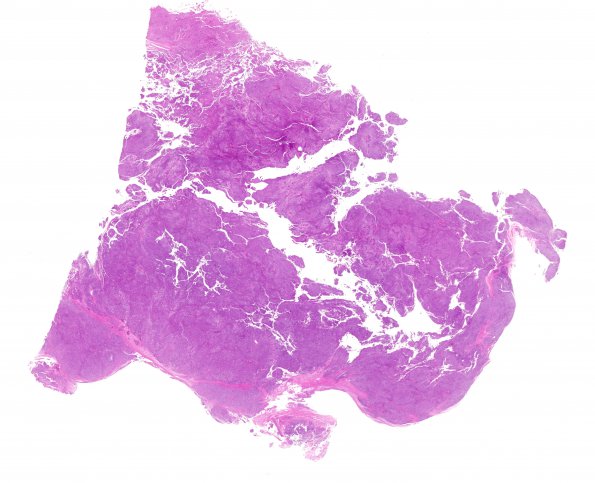
Washington University Experience | NEOPLASMS (MENINGIOMA) | Anaplastic | 27A1 Meningioma, anaplastic, gland meta (Case 27) H&E WM
Case 27 History ---- The patient had a prior medical history of a meningeal tumor that was first operated upon in 2001 with subsequent recurrences in 2003 and 2006. Original and first recurrence were both diagnosed as meningioma. Molecular genetic studies on the 2006 specimen are negative for the t(X;18) characteristic of synovial sarcoma. ---- 27A1-6 Microscopic sections reveal an anaplastic meningioma with glandular metaplasia, WHO grade III. Tumor cell density is high in this solid neoplasm that primarily displays a sheeting architecture, but in addition exhibits definitive gland formation. These glands are scattered throughout the tumor and are generally formed by a single layer of cells. Occasionally, micropapillary structures emanate from the glandular epithelium. Clusters of less well defined epithelioid differentiation are also seen. Focally, tumor cells are arranged in single files. There is a focal suggestion of mini-whorl formation, but otherwise better differentiated meningothelial-type tumor features are absent. The majority of tumor cells bear a small amount of ill-defined eosinophilic cytoplasm. Associated tumor nuclei are somewhat monotonous in morphology, being oval in shape while exhibiting relatively open chromatin and prominent nucleoli. The cytologic appearance of the glandular and interglandular tumor cells is very similar. There are focal clear cell features, but pseudopsammoma bodies are absent. Mitoses are numerous and include abnormal forms; mitoses are quantified at 30/10HPF.

