Table of Contents
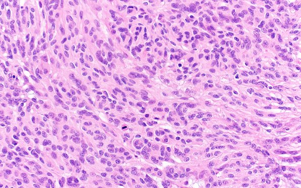
Washington University Experience | NEOPLASMS (MENINGIOMA) | Anaplastic | 6A1 Meningioma, anaplastic (Case 6) B3 H&E40X
Case 6 History ---- The patient was a 64-year-old woman with 6-12 months of intermittent aphasia and alexia, and two years of balance difficulties. Brain MRI showed a likely extra-axial, predominantly homogenously enhancing mass abutting the left tentorial leaflet and extending into the left temporal and parietal lobes with surrounding vasogenic edema. Operative procedure: Frameless stereotactic image-guided left posterior temporal craniotomy for resection of dural-based mass. ---- 6A1-4 This meningothelial neoplasm exhibits multiple growth patterns, including whorl formation and fascicular growth featuring herringbone and pseudo-Verocay growth patterns. The tumor cells have ovoid nuclei, coarse-to-vesicular chromatin, prominent nucleoli, and moderate amounts of eosinophilic cytoplasm with indistinct cell borders. Mitotic activity is high (>23 mitoses/10HPF). The tumor shows focal brain invasion, sheeting architecture, macronucleoli, large areas of spontaneous necrosis, and hypercellularity. (H&E)

