Table of Contents
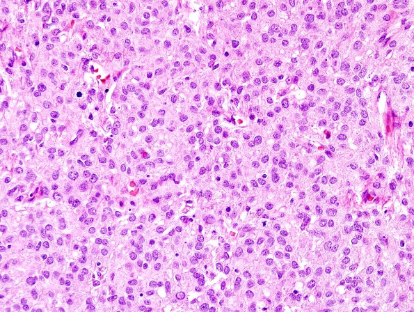
Washington University Experience | NEOPLASMS (MENINGIOMA) | Anaplastic | 7B3 Meningioma, anaplastic (Case 7) H&E 6.jpg
Much of the tumor tissue appears in hypercellular sheets, with multifocal spontaneous necrosis, sparse foci of 'small cell change,' and a few areas exhibiting clear cell pattern. Mitotic activity is brisk in papillary areas, ranging up to at least 22 per 10 contiguous high power (40x objective) fields. Adherent gliotic brain tissue is noted in several areas and, in some areas, brain invasion.

