Table of Contents
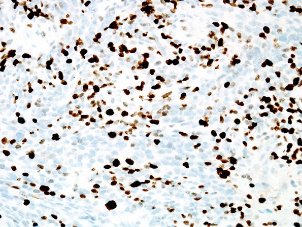
Washington University Experience | NEOPLASMS (MENINGIOMA) | Anaplastic | 7D Meningioma, anaplastic (Case 7) Ki67 2.jpg
Immunohistochemically stained sections show nuclear reactivity for proliferation marker Ki67 (MIB-1 antibody) in sheeted areas ranging up to at least 15%, and in papillary areas ranging up to approximately 40%. ---- Ancillary data (not shown): Reactivity for GFAP highlights adherent brain parenchyma with and without areas of unequivocal invasion. ---- FISH studies show evidence for loss of the p16/CDKN2A locus at 9p21. ---- Comment: This histological/immunohistochemical/genetic pattern supports the diagnosis 'anaplastic meningioma, WHO grade III.' Anaplasia is supported by high mitotic index (exceeding a threshold of 20/10HPF and widespread papillary histological pattern. Loss of 9p21 is found in most anaplastic meningiomas, and is associated with relatively unfavorable prognosis.

