Table of Contents
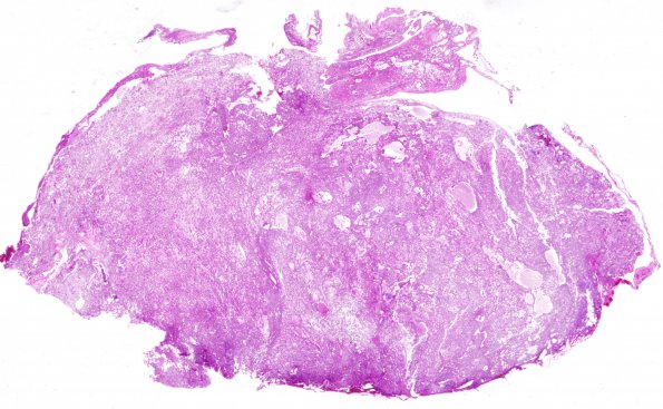
Washington University Experience | NEOPLASMS (MENINGIOMA) | Angiomatous | 10C1 Meningioma, angiomatous (Case 10) H&E WM
10C1-6 Sections show a highly vascular, moderately cellular neoplasm. The majority of blood vessels are markedly hyalinized. The tumor shows moderate nuclear pleomorphism, as well as scattered bizarre tumor nuclei. The tumor cells have abundant clear eosinophilic cytoplasm. Scattered nuclear holes and pseudoinclusions are also evident. There are areas with microcystic change and some tumor cells have a foamy xanthomatous appearance. Focal features more consistent with a typical meningioma with whorls are seen but there are no definite psammoma bodies. The nuclear atypia is most likely degenerative in nature, given the lack of appreciable mitotic activity. A single section shows a small fragment of brain parenchyma, with sharp demarcation from the adjacent tumor.

