Table of Contents
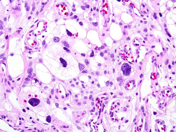
Washington University Experience | NEOPLASMS (MENINGIOMA) | Angiomatous | 10C6 Meningioma, Micro Angio (Case 10) 2.jpg
Additional illustrative image.(H&E). ---- Ancillary data (not shown): There was patchy positivity for epithelial membrane antigen (EMA) and strong nuclear immunoreactivity for progesterone receptor in a subset of tumor cells. There were scattered CD163 positive cells, consistent with a contribution of an infiltrate of macrophages, although tumor cells are mostly negative. The morphologic and immunohistochemical features are consistent with the diagnosis of meningioma, angiomatous/microcystic variant, WHO grade I.

