Table of Contents
Washington University Experience | NEOPLASMS (MENINGIOMA) | Angiomatous | 12A1 Meningioma (Case 12)
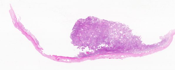
Case 12 History ---- The patient was a 52 year old woman who presented with history of headaches and deteriorating memory. On imaging, she was found to have a large left falcine mass and another satellite lesion in the right parietal region. She underwent a resection of left falcine mass in February of 2011 that was diagnosed as a papillary meningioma, and now here for resection of satellite lesion that was being followed-up. Operative procedure: Right parietal craniotomy for tumor resection. ---- 12A1-4 Attached to a portion of dura mater is a heterogeneous appearing cellular tumor with sheeted arrangements of cells interrupted by numerous thick-walled vessels and focal formation of whorls. At low magnification the tumor on low power has a zonation pattern with pale and dark areas. In the pale areas, the tumor cells have moderate amount of eosinophilic cytoplasm with prominent pseudoinclusions in the nuclei. In the darker areas, the cells appear much smaller and have scant amount of cytoplasm. Prominence of nucleoli is noted in multiple areas and mitoses are scattered (~4/10 hpf). Focal lipomatous change in the tumor cells, patchy degenerative atypia and rare psammoma bodies are also present.

