Table of Contents
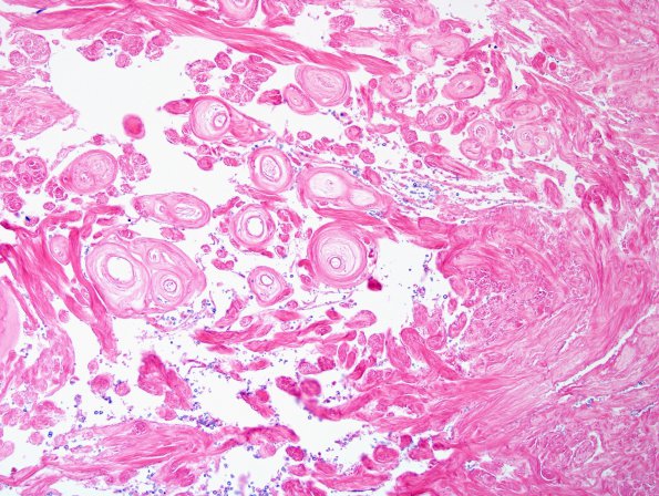
Washington University Experience | NEOPLASMS (MENINGIOMA) | Angiomatous | 13B2 Meningioma (DDx SFT) (Case 13) H&E 7.jpg
13B2-6 Sections show meningioma composed of islands of meningothelial cells intercalated between collagenous bands and large numbers of hyalinized blood vessels. Tumor cells are meningothelial with indistinct cell borders and occasionally fibroblastic with small bland appearing nuclei and rare mitotic figures. There are no significant areas of small cell change, hypercellularity, macronucleoli, or brain invasion. Hyalinized vessels vary markedly in size and are highlighted by trichrome and pentachrome stains.

