Table of Contents
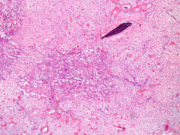
Washington University Experience | NEOPLASMS (MENINGIOMA) | Angiomatous | 15A1 Meningioma, Angiomatous & Xanthomatous (Case 15) H&E 5
Case 15 History ---- The patient was a 38 year old woman with a history of seizures. Imaging studies revealed a 4.6 x 2.9 cm enhancing right fronto-parietal parafalcine mass. This was associated with minimal mass effect and was clinically felt to be most suggestive of meningioma.---- 15A1-5 Sections reveal a highly vascular, moderately cellular neoplasm. The majority of blood vessels are markedly hyalinized. The tumor shows moderate nuclear pleomorphism, as well as scattered bizarre tumor nuclei. The tumor cells have abundant clear foamy to eosinophilic cytoplasm. Scattered nuclear holes and pseudoinclusions are also evident. Only vague whorls are seen and there are no definite psammoma bodies. The nuclear atypia is most likely degenerative in nature, given the lack of appreciable mitotic activity.

