Table of Contents
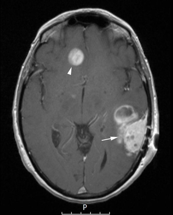
Washington University Experience | NEOPLASMS (MENINGIOMA) | Angiomatous | 2A1 Meningioma, angiomatous (Case 2) T1W copy - Copy
Case 2 History ---- The patient was a 67 year old woman who presented with headache and a recent history of memory and word-finding difficulty. At an OSH, brain imaging showed a heterogeneously enhancing 3.5 x 3 x 2.5 cm dura associated mass with internal non-enhancing cysts in the left parietal lobe, an adjacent rim-enhancing 3 x 2.5 cm lesion in the deeper white matter, and a spheroidal ~1.5 cm rim-enhancing mass in the right frontal lobe with associated edema. Biopsy of the superficial portion of the right frontal lobe lesion was performed at outside hospital and showed meningioma, WHO grade I. Operative procedure: Stealth-guided craniotomy for resection of brain tumor with intraoperative MRI. ---- 2A1,2 MRI confirmed an enhancing extra-axial mass in the right frontal lobe (arrowhead, 2A1) and an intrinsic left parietal mass lesion (arrow, 2A1) proven to represent coexistence of meningioma and a high grade glioma as shown in T1-weighted (2A1) and T2-weighted (2A2) contrast administered scans.

