Table of Contents
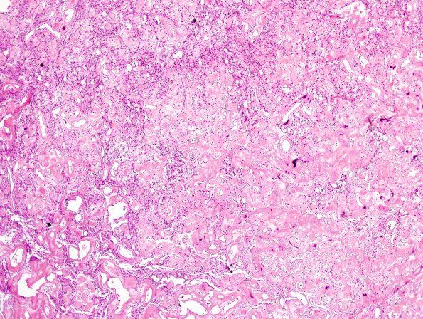
Washington University Experience | NEOPLASMS (MENINGIOMA) | Angiomatous | 2B2 Meningioma, (Case 2) H&E 3.jpg
2B2-7 This tumor represents an angiomatous meningioma with lesser microcystic, lipomatous, and meningothelial features. Hyalinized blood vessels with thickened walls and incipient or complete focal mural calcifications predominate, but are intermixed with sheets and lobules of meningothelial cells in many areas. Many of the tumor cells have round to oval nuclei with some variation in size, small nucleoli, occasional intranuclear clearings, moderate eosinophilic cytoplasm, and indistinct cell borders. Many others have lucent cytoplasm or are filled with numerous clear vesicles. A few psammoma bodies are noted. Atypical features of hypercellularity, sheeting, small cell change, macronucleoli, and 'spontaneous' necrosis are not observed. Mitotic figures are rare (<1/10HPF). There is no evidence of brain invasion.

