Table of Contents
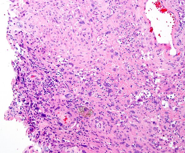
Washington University Experience | NEOPLASMS (MENINGIOMA) | Angiomatous | 3B1 Meningioma, angiomatous (Case 3) H&E-3
3B1-5 Sections show a remarkably vascular meningothelial neoplasm with marked degenerative atypia. The neoplastic cells show focal prominence of nucleoli. However, other features like hypercellularity, sheeting, small-cell change or necrosis are not seen. Mitoses are rare (2/10HPF). There is collagen deposition and rare psammoma bodies. Patchy areas of lymphoplasmacytic cell infiltrate and scattered hemosiderin deposits are noted.

