Table of Contents
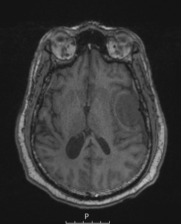
Washington University Experience | NEOPLASMS (MENINGIOMA) | Angiomatous | 4A1 Meningioma, angiomatous (Case 4) T1noC - Copy
Case 4 History ---- The patient was a 61-year-old man with a history of high grade cerebellar meningioma status post resection in August 2019. He presents with 4.7 cm left frontal dural-based homogenously enhancing mass consistent with a meningioma. Operative procedure: Left frontal craniotomy for tumor resection. ---- 4A1-3 MRI examination of a left frontal mass as shown with T1-weighted without (4A1) and with contrast (4A2) and T2-weighted contrast administered (4A3) scans.

