Table of Contents
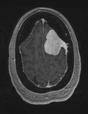
Washington University Experience | NEOPLASMS (MENINGIOMA) | Angiomatous | 5A2 Meningioma, angiomatous and microcystic (Case 5) T1W - Copy
MRI studies show a hyperintense mass with T1-weighted contrast enhanced scan (note the dural tail) and T2-weighted image with contrast (5A3).

