Table of Contents
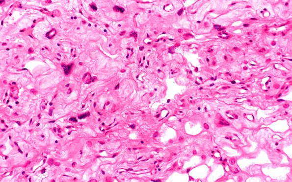
Washington University Experience | NEOPLASMS (MENINGIOMA) | Angiomatous | 5B4 Meningioma, angiomatous and microcystic (Case 5) H&E 2
Much of the tumor tissue is formed by rather pleomorphic epithelioid cells with variably moderate to abundant pale eosinophilic or vacuolated cytoplasm, distinct cell borders, and nuclei that range from small and oval to very large and hyperchromatic (consistent with 'degenerative atypia'). Interspersed are numerous hyalinized thick-walled blood vessels of small to large caliber, and microcysts often filled with flocculent eosinophilic material.

