Table of Contents
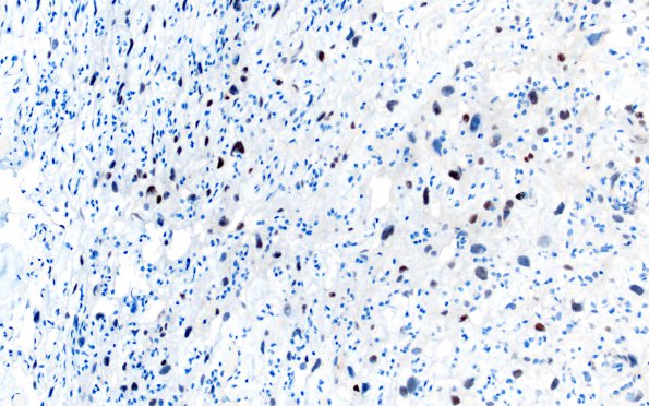
Washington University Experience | NEOPLASMS (MENINGIOMA) | Angiomatous | 5D Meningioma, angiomatous and microcystic (Case 5) PR 1
5D Immunohistochemistry reveals strong reactivity for progesterone receptor in a minority of tumor cell nuclei, with some preferential staining of larger nuclei. ---- Ancillary studies (not shown): Reactivity for proliferation marker Ki-67 (MIB-1 antibody) stains a regionally variable proportion of tissue nuclei; generally, the proliferation index is very low, but it ranged focally up to 4.9%. The radiological, histomorphologic and immunohistochemical features of this lesion are consistent with a meningioma with angiomatous and microcystic features, WHO Grade I. As part of this evaluation, slides from the patient's partial nephrectomy specimen were reviewed; the patient's brain tumor and papillary renal cell carcinoma are histomorphologically distinct.

