Table of Contents
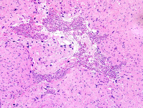
Washington University Experience | NEOPLASMS (MENINGIOMA) | Angiomatous | 6A1 Meningioma, angiomatous, xanthomatous (Case 6) H&E 5.jpg
Case 6 History ---- The patient was an 81 year old woman with a 5.2 cm enlarging right frontal parafalcine extra-axial mass causing cerebral vasogenic edema and midline shift. This lesion was followed for 2 years before she began to experience dizziness, personality changes and seizures. Operative procedure: Resection. ---- 6A1-7 Sections of the resected intracranial mass show a dural-based tumor with extensive hyalinization and marked degenerative atypia (ancient change). Tumor cells are not obviously organized into whorls. Accumulation of clustered small vessels and large vessels in combination with hyalinization provides an angiomatous appearance. Xanthomatous individual cells are noted. Mitotic activity is not detected, brain invasion is absent and there are no additional atypical features (e.g., spontaneous necrosis).

