Table of Contents
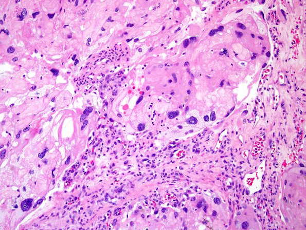
Washington University Experience | NEOPLASMS (MENINGIOMA) | Angiomatous | 6A4 Meningioma, angiomatous, xanthomatous (Case 6) H&E 6.jpg
Sections of the resected intracranial mass show a dural-based tumor with extensive hyalinization and marked degenerative atypia (ancient change). Tumor cells are not obviously organized into whorls. Accumulation of clustered small vessels and large vessels in combination with hyalinization provides an angiomatous appearance. Xanthomatous individual cells are noted.

