Table of Contents
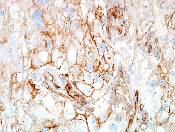
Washington University Experience | NEOPLASMS (MENINGIOMA) | Angiomatous | 6M Meningioma, angiomatous, xanthomatous (Case 6) CD99 1.jpg
CD99 shows faint tumor cell reactivity ---- Ancillary data (not shown): Glial fibrillary acidic protein shows no evidence of brain invasion. Pancytokeratin, desmin, synaptophysin and Melan-A are negative. Synaptophysin is completely negative.
Diagnosis Comment
This tumor is unusual in multiple ways. Most obvious is the widespread presence of bizarre nuclear morphology, which, in the absence of mitotic figures or elevated proliferative activity, is likely to indicate a degenerative phenomenon. Immunoreactivity for markers typically associated with meningothelial differentiation, including progesterone receptor and epithelial membrane antigen, is sparse but unequivocally positive. Overall, we believe the radiography, histomorphology and immunohistochemistry profile are most compatible with angiomatous meningioma without atypical features or other findings to warrant a grade above WHO grade I.

