Table of Contents
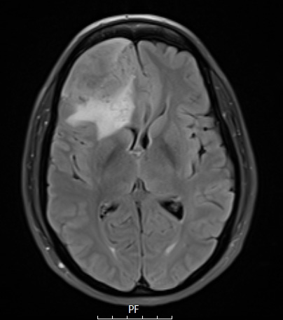
Washington University Experience | NEOPLASMS (MENINGIOMA) | Angiomatous | 8A1 Meningioma, angiomatous (Case 8) TIRM - Copy
Case 8 History ---- The patient is a 49–year–old woman with history of headache and brain MRI demonstrating a large dural based lesion involving the right frontal and right anterior fossa regions with secondary mass effect, that represent meningioma. Operative procedure: Craniotomy with right frontal tumor resection. ---- 8A1-3 MRI studies show a right parafalcine mass viewed in TIRM (8A1), T1- (8A2) and T2-weighted contrast enhanced (8A3) scans.

