Table of Contents
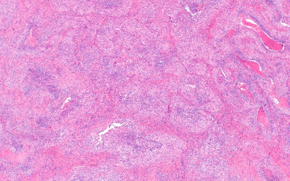
Washington University Experience | NEOPLASMS (MENINGIOMA) | Angiomatous | 8B1 Meningioma, sclerotic (Case 8) H&E 4X (2)
8B1-3 H&E stained tumor cells have ovoid nuclei, with focal areas that have prominent nucleoli, intranuclear clearing and pseudoinclusions, and moderate amounts of eosinophilic cytoplasm. Most of the tumor cells have indistinct cell borders and are arranged in whorls and fascicles. Mitotic figures are readily identified up to 5/10HPF. There are also numerous areas of necrosis seen in several blocks. Also there is eosinophilic material throughout the resection specimen which is positive for trichrome, confirming fibrosis and negative for amyloid deposition. Many areas have an angiomatous appearance.

