Table of Contents
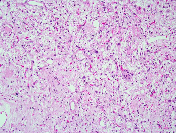
Washington University Experience | NEOPLASMS (MENINGIOMA) | Angiomatous | 9B1 Meningioma, Microcystic Angiomatous (Case 9) 1.jpg
9B1,2 Sections reveal a low to moderately cellular solid-appearing neoplasm with rich vascularity and abundant loose microcystic stroma. There is moderate nuclear pleomorphism, with the majority of tumor cells containing oval nuclei, delicate chromatin, and thin wispy cytoplasmic processes. Scattered bizarre nuclei are also encountered and are felt to represent degenerative atypia, given their relative lack of association with increased mitotic activity. There are also occasional nests of tumor cells with a more epithelioid morphology, resembling meningothelial cells. Many of the blood vessels are markedly hyalinized. No tumor necrosis is evident.

