Table of Contents
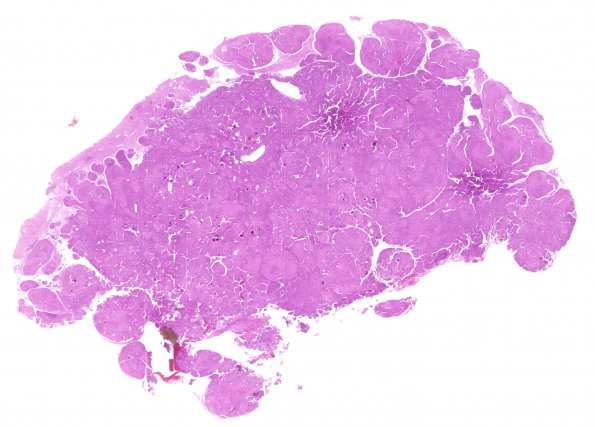
Washington University Experience | NEOPLASMS (MENINGIOMA) | Brain Invasion | 10A1 Meningioma, brain invasion (Case 10) H&E WM
Case 10 History ---- The patient was a 38-year-old woman who experienced a seizure during delivery of her first child. At that time, neuroimaging work up demonstrated an intracranial mass. Recent MRI showed a bifrontal 3.5 cm extra-axial, post-contrast enhancing skull base mass in the area of the olfactory groove. Operative procedure: Resection. ---- 10A1-3 This meningothelial neoplasm has a solid growth pattern and is arranged in lobules and whorls with prominent brain invasion. Necrosis and other atypical features are not identified. The tumor cells are syncytial without distinct cell borders, and they have abundant eosinophilic cytoplasm. Mitotic figures are infrequent (<1/10HPF).

