Table of Contents
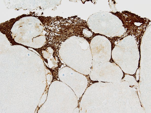
Washington University Experience | NEOPLASMS (MENINGIOMA) | Brain Invasion | 10C2 Meningioma, brain invasion (Case 10) GFAP 1.jpg
GFAP immunoreactivity highlights the multifocal areas of tumor invasion into the brain parenchyma. (GFAP IHC) ---- The proliferation marker Ki67 stains a subset of tumor cell nuclei at variable density, ranging focally up to 4.6%.

