Table of Contents
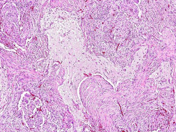
Washington University Experience | NEOPLASMS (MENINGIOMA) | Brain Invasion | 12A1 Meningioma, brain invasion (Case 12) H&E 2.jpg
This tumor is represented by a neoplastic proliferation of meningothelial cells arranged in whorls, nests, and vaguely fascicular architecture admixed with chronic inflammation. Mitotic figures are not identified. No subjective atypical features, including hypercellularity, small cell change, necrosis, or sheeted architecture are observed. Brain invasion is characterized by small islands of glial tissue surrounded by meningothelial cells. This tumor is best classified as a meningioma with brain invasion, WHO grade II which was virtually identical in histomorphologic appearance to that seen in the original 2009 specimen.

