Table of Contents
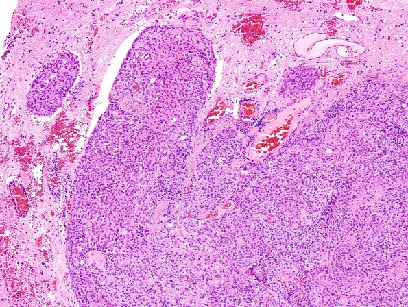
Washington University Experience | NEOPLASMS (MENINGIOMA) | Brain Invasion | 14A Meningioma, brain invasion WHO-II (Case 14) H&E 6
Case 14 History ---- The patient was a 62 year old woman with a history of a focal motor seizure. MRI of the brain showed a 5.4 x 3.9 cm extra-axial contrast-enhancing mass centered in the left precentral and postcentral gyrus. Operative procedure: Craniotomy with resection of lesion. ---- 14A Sections show a hypercellular meningothelial neoplasm with both nodular and sheet-like areas of growth. The neoplastic cells have round to ovoid shaped nuclei with mildly stippled chromatin, prominent macronucleoli, and intranuclear cytoplasmic 'pseudo'-inclusions. Portions of the lesion show small cell change. Mitotic figures number up to 8/10HPF. A portion of the lesion shows adherent brain parenchyma and brain invasion.

