Table of Contents
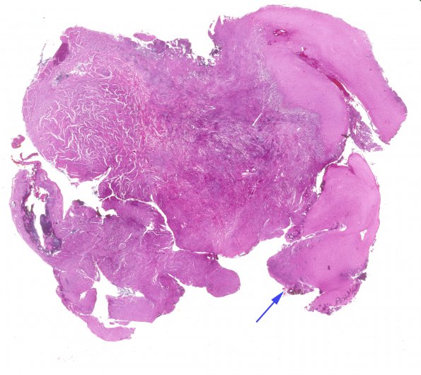
Washington University Experience | NEOPLASMS (MENINGIOMA) | Brain Invasion | 17A1 Meningioma, rhabdoid & brain invasion (Case 17) H&E WM
Case 17 History ---- The patient was a 26 year old man who underwent two resections for brain tumors at the University of Missouri in Columbia, one in January (ganglioglioma) and the second in October of 2007 (rhabdoid meningioma). ---- 17A1-4 One component has a low mitotic index and in some areas, closely resembles meningioma. The majority of the second tumor appears much more cellular and varies from intersecting fascicles of relatively bland-appearing spindled cells with scattered whorl formation to large, discohesive sheets of pleomorphic, rhabdoid cells with vesicular nuclei, prominent nucleoli, and eccentric bellies of eosinophilic cytoplasm. The latter component shows a high mitotic index and large zones of tumor necrosis.

