Table of Contents
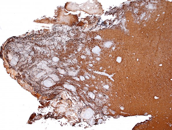
Washington University Experience | NEOPLASMS (MENINGIOMA) | Brain Invasion | 17B2 Meningioma, rhabdoid & brain invasion (Case 17) GFAP 4X.jpg
The GFAP stain highlights reactive astrocytes in the adjacent brain parenchyma and islands of infiltrating tumor. The area shown in 17A1 (arrow) is the same area shown in 17B2,3 with tumor invasion.

