Table of Contents
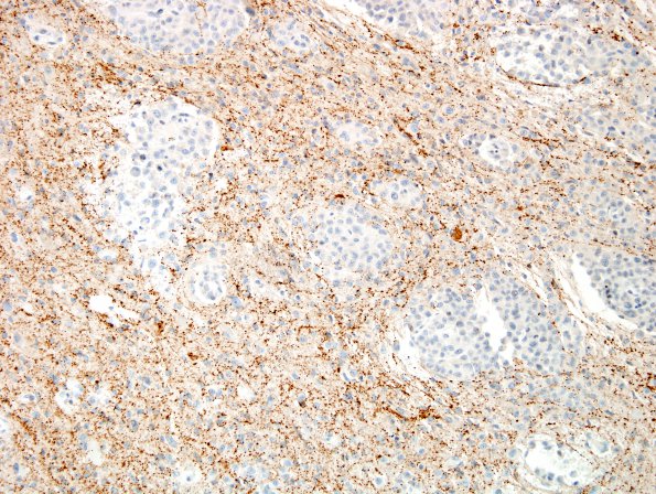
Washington University Experience | NEOPLASMS (MENINGIOMA) | Brain Invasion | 17D2 Meningioma, rhabdoid & brain invasion (Case 17) SYN 1.jpg
Nodules of tumor displace nearby neurofilament containing axons (17D1) and synaptophysin immunoreactive elements (17D2). ---- Additional results (not shown): An immunostain for EMA shows patchy positivity, particularly in more meningothelial-appearing regions. The vimentin stain is diffusely positive and highlights the rhabdoid morphology. Only rare chromogranin positive neurons are seen at the adjacent brain. The tumor cells are negative for cytokeratin and actin. A repeat EMA stain highlights predominantly the meningothelial-appearing regions of the tumor, although some of the rhabdoid cells are also positive. A stain for INI-1 (BAF47) reveals retained nuclear immunoreactivity within tumor cells, essentially excluding the possibility of atypical teratoid/rhabdoid tumor. FISH showed polysomy 22 but this genetic pattern is considered non-specific. ---- Comment: The morphologic and immunohistochemical features are consistent with the diagnosis of rhabdoid meningioma, WHO grade 3 in the second resection specimen.

