Table of Contents
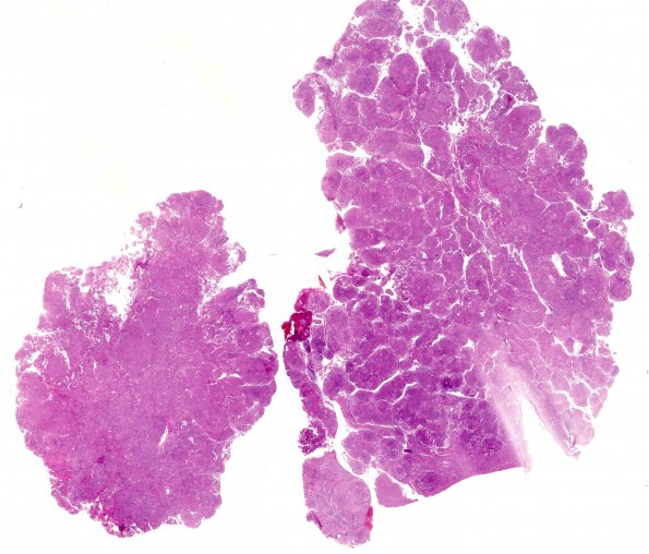
Washington University Experience | NEOPLASMS (MENINGIOMA) | Brain Invasion | 18A1 Meningioma, WHO II brain invasion (Case 18) H&E WM
Case 18 History ---- The patient was a 35-year-old woman with an incidentally discovered anterior falcine lesion suspicious for a meningioma following a car accident during which she suffered a concussion. A head CT scan showed no hemorrhage but did demonstrate a frontal mass. MRI showed a 2.9 x 2.7 x 2.3 cm homogeneous enhancing right olfactory groove meningioma. Operative procedure: Craniotomy for meningioma. ---- 18A1-4 Sections of the resected meningioma show groups of neoplastic meningothelial cells separated by fibrous septa. Architecturally, neoplastic cells are arranged in tight concentric whorls and also in less compact sheets of meningothelial cells. Cytologically, tumor cells display frequent nuclear grooves, holes and pseudoinclusions.

