Table of Contents
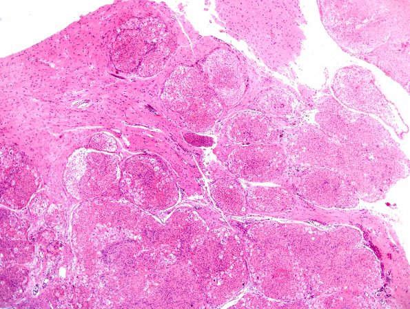
Washington University Experience | NEOPLASMS (MENINGIOMA) | Brain Invasion | 18A4 Meningioma, WHO II brain invasion (Case 18) H&E 8.jpg
Sections of the resected meningioma show groups of neoplastic meningothelial cells separated by fibrous septa. Architecturally, neoplastic cells are arranged in tight concentric whorls and also in less compact sheets of meningothelial cells. Cytologically, tumor cells display frequent nuclear grooves, holes and pseudoinclusions.

