Table of Contents
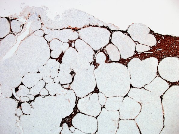
Washington University Experience | NEOPLASMS (MENINGIOMA) | Brain Invasion | 18B Meningioma, WHO II brain invasion (Case 18) GFAP 3.jpg
Multifocal invasion deep into the brain parenchyma is confirmed with GFAP immunoreactivity. (GFAP IHC) ---- There is multifocal spontaneous tumor necrosis, however other atypical histological features are focal or absent. Mitotic activity is rare. ---- A panel of immunohistochemical stains was performed to additionally characterize this neoplasm. Progesterone receptor is diffusely and strongly positive in tumor cell nuclei. Ki-67 is focally elevated to give a maximum proliferation index of 13%. These findings are diagnostic of a WHO Grade 2 meningioma due to the presence of unequivocal brain invasion.

