Table of Contents
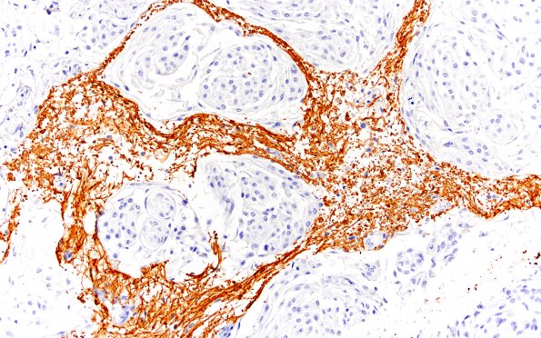
Washington University Experience | NEOPLASMS (MENINGIOMA) | Brain Invasion | 1B3 Meningioma, WHO II, brain invasion (Case 1) N 20X GFAP
Higher magnification images of tumor nodules separated by intercalated gliotic brain parenchyma. (GFAP IHC) ---- These histomorphologic and immunohistochemical features are consistent with the diagnosis of atypical meningioma with brain invasion, WHO grade 2. A Ki67 immunostain demonstrates a proliferation index of 3% with focal elevation to 5%. A subset of tumor cells showed weak PR nuclear immunoreactivity.

