Table of Contents
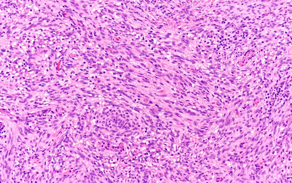
Washington University Experience | NEOPLASMS (MENINGIOMA) | Brain Invasion | 21B1 Meningioma, brain invasion (Case 21) H&E 20X 3
21B1-3 Hematoxylin and eosin stained sections show a proliferation of neoplastic meningothelial cells arranged in syncytial lobules and spindled elements. Tumor cells have ovoid nuclei, coarse chromatin, nuclear clearing, nuclear pseudoinclusions, nuclear grooves, and eosinophilic cytoplasm with indistinct cell borders. Mitoses were rare (<1/10HPF). There were macronucleoli, focal areas of necrosis, and focal small cell change. (H&E)

