Table of Contents
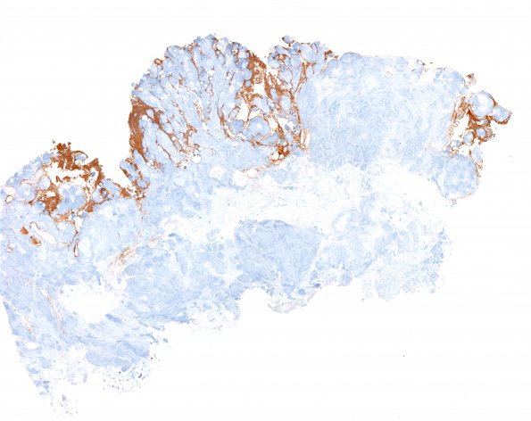
Washington University Experience | NEOPLASMS (MENINGIOMA) | Brain Invasion | 2A1 Meningioma (Case 2) brain invasion GFAP 1
Case 2 History ---- The patient was a 48 year old man with a left frontal dura-based mass and a history of seizures. The tumor was embolized a few days before surgery. ---- The tumor cells have vesicular, intranuclear pseudoinclusions, focal small cell change, macronuclei, sheeting and scattered mitoses (<3/10HPF). In addition, some of the tumor cells also demonstrate macronucleoli. These histomorphologic features are diagnostic of an atypical meningioma, WHO grade II. ---- 2A1-3 Long stretches of meningioma infiltrate between gliotic brain tissue forming surrounded islands (GFAP IHC)

