Table of Contents
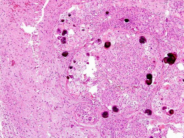
Washington University Experience | NEOPLASMS (MENINGIOMA) | Brain Invasion | 4A1 Meningioma & brain invasion (Case 4) H&E 5.jpg
Case 4 History ---- The patient was a 55-year-old woman with a left frontal dural-based mass that was incidentally diagnosed in 2009 following a motor vehicle accident. For the last 3 years of she experienced weakness, increased dizziness and blurry vision. Operative procedure: Tumor excision. ---- 4A1-3 Multiple images show a meningothelial neoplasm that is widely brain-invasive. The tumor is otherwise benign-appearing and arranged predominantly in cellular whorls with abundant psammoma bodies. Mitotic figures are <1/10 HPF. Atypical features such as spontaneous necrosis, sheeting, macronucleoli, or hypercellularity are not identified. A few foci of small cell change are noted. The tumor was diagnosed as an atypical meningioma with brain invasion, WHO Grade 2.

