Table of Contents
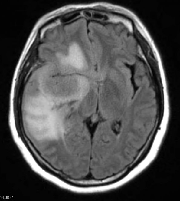
Washington University Experience | NEOPLASMS (MENINGIOMA) | Brain Invasion | 5A1 Meningioma, atypical, brain invasion (Case 5) FLAIR 2 - Copy
Case 5 History ---- The patient was a 56 year old woman who was status-post hysterectomy in 2004 which identified poorly differentiated adenocarcinoma of the endocervix and a separate focus of well-differentiated adenocarcinoma of the uterus (endometrioid type, FIGO grade I, without myometrial invasion). One year prior to surgery she presented with increasing tremors on her left side for several months, and diplopia. ---- 5A1-4 MRI revealed a 4.8 x 4.8 x 4.6 extra-axial, homogenously-enhancing mass centered within the right middle cranial fossa, anterior to the temporal lobe. ---- 5A1 The tumor mass and surrounding edema is well seen in this FLAIR image.

