Table of Contents
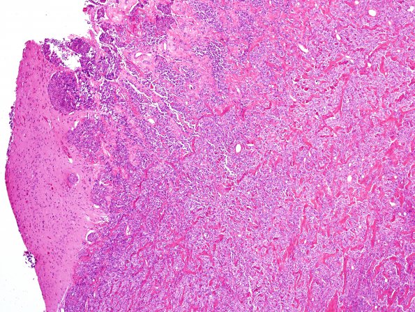
Washington University Experience | NEOPLASMS (MENINGIOMA) | Brain Invasion | 6A2 Meningioma, brain invasion (Case 6) H&E 4.jpg
The neurosurgical resection specimens show a hypercellular meningothelial neoplasm with areas of sheet-like 'patternless' growth, areas of necrosis, prominent nucleoli, and multifocal areas of brain invasion.

