Table of Contents
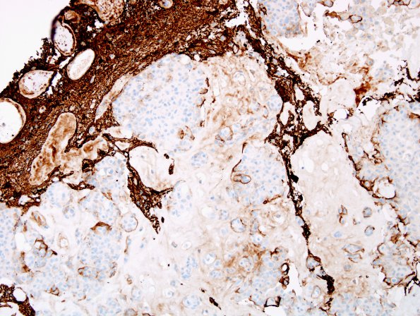
Washington University Experience | NEOPLASMS (MENINGIOMA) | Brain Invasion | 6E3 Meningioma, brain invasion (Case 6) GFAP 3.jpg
GFAP immunoreactivity highlights the multifocal areas of brain invasion by the tumor. (GFAP IHC) ---- The proliferation marker Ki67 stains a small subset of tumor nuclei, ranging up to 1.2%. ---- Comment: The overall histomorphologic and immunophenotypic characteristics describe an atypical meningioma with brain invasion, WHO grade 2. The designation of 'atypical' is applied in this case because of the multiple subjective criteria present including hypercellularity, sheet-like 'patternless' growth, prominent nucleoli, and necrosis. The mitotic count is quite low (<1/10 high powered fields), which is reflected in the low Ki67 proliferation index (1.2%). Either atypical features or brain invasion, independent of one another, are sufficient to grade a meningioma as WHO grade 2.

