Table of Contents
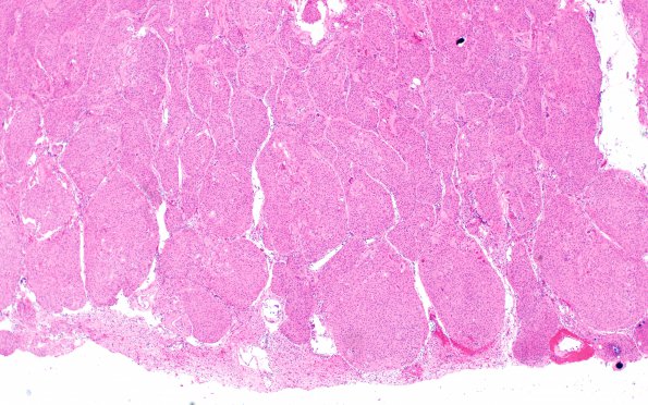
Washington University Experience | NEOPLASMS (MENINGIOMA) | Brain Invasion | 9A1 Meningioma, brain invasion (Case 9) H&E 4X
Case 9 History ---- The patient was a 60-year-old woman presenting with episodes of confusion. Imaging showed a solid enhancing dural-based extra-axial mass arising from the right sphenoid near the vertex, likely represent meningioma. Operative procedure: Right frontotemporal craniotomy for tumor resection. ---- 9A1-4 H&E stained tissue shows a neoplasm arranged in lobules. The cells have a syncytial pattern with indistinct cell borders and abundant eosinophilic cytoplasm. No mitoses are identified. Psammoma bodies are present. Brain invasion is identified with areas of reactive Rosenthal fibers, seen best in image #9A3.

